
Crognale Lab's research focuses on the physiological basis for visual and other sensory processing and attention. His research spans all levels of sensory processing from the photoreceptors to higher cortical functions such as attention and human factors. His major contributions come in the areas of comparative visual processing, visual development and aging, genetics of color vision, and applications of human electrophysiology to the study of visual function.
The efforts from his lab include application of high density EEG, electroretinography (ERG), functional near infrared spectroscopy (fNIRS), fMRI and adaptive optics scanning laser ophthalmoscopy (AOSLO) to questions of visual function.
Attentional modulation of Visual Evoked Potential (VEP) response to black and white checkerboard
patterns is readily observed, as reported in prior literature. However, results from more recent studies
have shown that chromatic VEP responses, known to reflect largely early cortical processing, are robust
to attentional modulation. We employed several levels
of difficulty of a distracting task, multiple object tracking (MOT), to insure sufficient attentional
modulation.
The VEP results support prior conclusions that chromatic onset VEP responses show little modulation with attentional shifts. We also replicated prior research that demonstrated strong attentional modulation of responses to standard achromatic (black and white) reversing checkerboard
stimuli. However, surprisingly, we also failed to observe a modulation of responses to the achromatic
onset grating stimuli, mirroring the results of the chromatic condition.
A well-known asymmetry exists between brightness induction for incremental and decremental targets, which is often characterized by the dogma that darkness induction is stronger than lightness induction. We explored this asymmetry with a simple brightness matching paradigm and discovered some novel quantitative properties. Observers adjusted the luminance of a matching disk to match its brightness to that of a fixed test disk surrounded by an annulus. The annulus luminance was experimentally varied to induce a brightness change in the test.
Average brightness matches were plotted against the annulus luminance on a log-log axis, the results from each condition were well-fit with a 2nd-order polynomial regression model. Increasing the annulus luminance increased the test brightness over one range (assimilation) and decreased it over another (contrast). We hypothesize that this pattern reflects the different underlying neural gains of ON and OFF cells in primate. This study is part of a collabration between Crognale Lab and Dr. Michael E. Rudd.

Color vision investigations often employ the concept of a color space based on a standard observer with standard cone fundamentals and cone ratios to calculate the relative activations of the cones and opponent mechanisms. However, since there are large variations in individuals’ cone fundamentals and luminosity function, each observer has their own unique color space.
To estimate the tilt of the equiluminant plane we employed a minimum motion task to stimuli modulated along the assumed opponent axes. To locate the individual opponent axes, we used an established adaptation/contrast-matching paradigm (Webster et al, 2000). For both tasks we included measurements from both the fovea and at 4-deg in the periphery wherein the values should vary significantly within an observer due to macular pigment and other retinal inhomogeneities. As predicted by our previous model, we found a strong correlation (r = .72, p=.003) between measures of luminance evidenced as a tilt in the equiluminant plane and estimates of the location of the SvsLM opponent axis revealed as a rotation within the equiluminant plane. This study is part of a collabration between Crognale Lab and Webster Lab.
The tritan line is the collection of colors that vary only in their stimulation of the S-cones. It is expected, then, that this line might be transformed in the visual periphery where macular pigment (which absorbs short-wavelength light) is minimal. We evaluate this tranformation at five levels of retinal eccentricity, using modified minimally distinct border and transient tritanopia tasks.
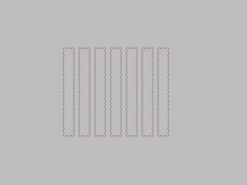
The watercolor effect is a visual illusion that manifests itself as a combination of long range color spreading and figure ground organization. The illusion is produced by the presence of adjacent edges of two contours that contrast each other in brightness, chromaticity, or a combination of both. We utilize a novel technique of measuring the cortical response of the illusion using the visual evoked potential (VEP).
The use of large fields in problematic to cone isolation due to retinal inhomogeneity. This series of studies evaluates novel large field stimuli in an attempt to account for changes across the retina.
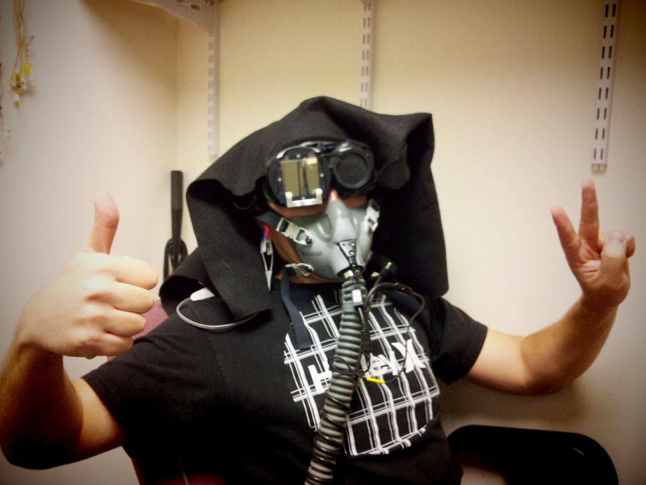
This study found that when blood oxygen saturation levels are reduced to around 75% under normobaric (normal air pressure) conditions, mild losses in color vision and more severe losses in recovery of sensitivity during dark adaptation occur.